Monday 20 February 2012

The Cerenkov Effect


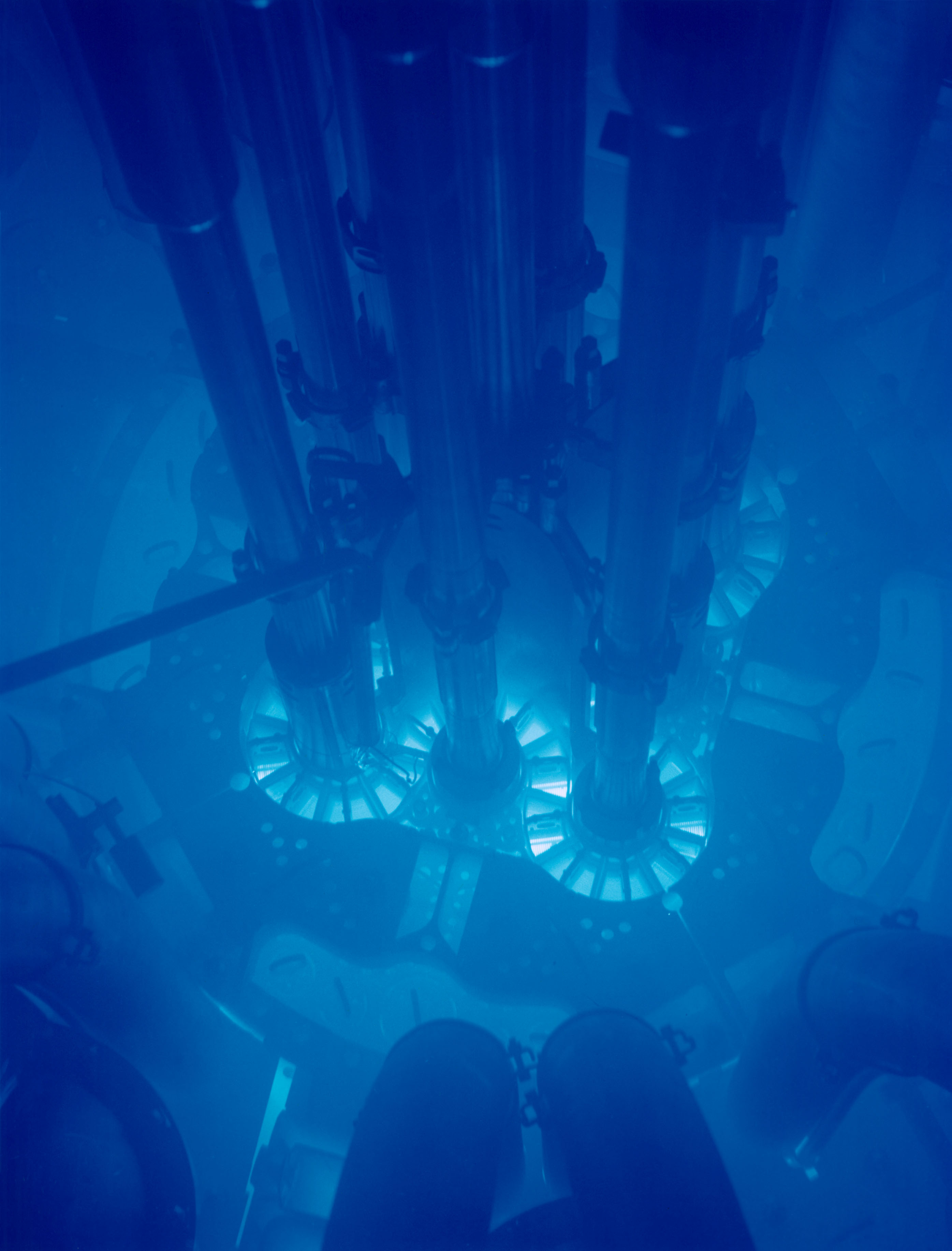
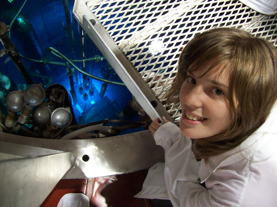
The Cerenkov effect is the light equivalent of a sonic boom (the noise heard when an object travels faster than the local speed of sound, e.g. the crack of a whip). When an object travels faster than the local speed of light (e.g. a high energy positron), a flash of blue light is emitted. This is the reason for nuclear reactors' eerie blue glow, and Watchmen character Doctor Manhattan's distinctive blue skin.


Cerenkov light is seen in nuclear reactors due to the nature of radioactive material they use. If the material decays by emitting a particle such as an electron or positron (Beta decay) as opposed to a photon (Gamma decay), chances are a large proportion of these particles will be traveling fast enough to satisfy the Cerenkov condition, i.e. faster than the speed of light within the radioactive material. This is why fuel rods and surrounding water (since the decay particles may be also traveling faster than the speed of light in water) appear to glow blue.


Friday 17 February 2012

A Little Background


Most of us will have heard the term 'PET Scan', even if we don't know what 'PET' stands for ('Positron Emission Tomography'), or what the scan actually entails. This video from the Helmholtz-Zentrum Dresden-Rossendorf research institute gives a good visual explanation of the process:


Basically, the patient ingests a radiotracer - a radioactive substance that is mixed with something else, such that the patient's body is 'tricked' into absorbing it as though it were a [useful] substance such as glucose. The radioactive part of the radiotracer is chosen such that (a) it emits positrons during decay and (b) it's half life - the time taken for it to decay to half its original amount - is a reasonable amount of time, e.g. an hour as opposed to 10,000 years.
Once the radiotracer is ingested, the body will transport it to the desired location (this depends on what particular radiotracer has been chosen). Positrons are then emitted in all directions into the surrounding tissue, and will annihilate with any electrons they come in contact with (this is simply what happens when matter and antimatter collide). This annihilation causes two photons (light particles) to be emitted in opposite directions. These are then detected by the PET camera on either side of the patient. 


This occurs many, many times, and a cross-section image of the body is then built up, based on the position where the photons hit the camera. The camera then moves up or down the body, allowing a three-dimensional image to be created.


PET Scans highlight areas of high uptake of particular chemicals - if a glucose analogue is chosen as the radiotracer, then the resulting image will show areas of high glucose uptake, i.e. metabolism. Higher than average metabolism is one of the key signs of cancer metastasis. This is why PET Scans are considered an important tool in medical diagnosis.